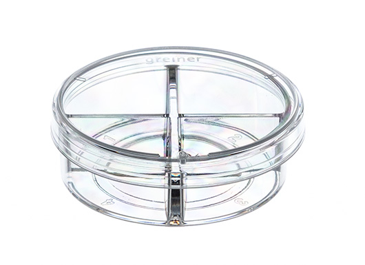
CELLview cell culture dish, four compartments, TC treated, Sterile, Case of 40
In Stock
Product code: 627870
MPN: 627870
Manufacturer: Greiner Bio-One
Shipping Weight: 0.77lbs (0.35kg)

Meet your sales rep Ben Morton, for the Philippines region.
Would you like to book 15 minutes on Ben Morton's calendar to get the best value for your budget?
Yes, book the timeCELLVIEW CELL CULTURE DISH, PS, 35/10 MM,GLASS BOTTOM, 4 COMPARTMENTS, TC, STERILE, 10 PCS./BAG
Description
Packaging
Detailed information
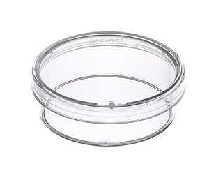
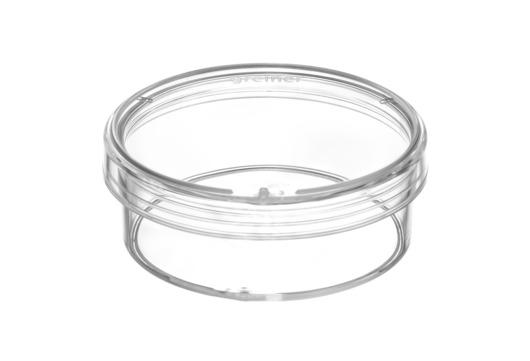
CELLview Cell Culture Dish with Glass Bottom
Drug treatment during live cell imaging
A multi-position time-lapse experiment was started and after acquiring six time points every two minutes drugs were added to the different wells as indicated:
| Description: |
CELLVIEW CELL CULTURE DISH, PS, 35/10 MM, GLASS BOTTOM, 4 COMPARTMENTS, TC, STERILE, 10 PCS./BAG |
|---|---|
| Sterile: | sterile |
| Packing unit: | 40 |
| Qty / inner pack: | 10 |
Packaging
| Packaging weight: | 0.35 kg |
|---|---|
| Packaging dimension: | 195 x 142 x 122 mm |
| Packing unit: | 40 |
| Qty / inner pack: | 10 |
| PAL: | 22800 |
Detailed information
| Filling volume (ml): | 0.00 |
|---|
CELLview Cell Culture Dish with Glass Bottom
- Free of detectable DNase, RNase, human DNA
- Non-pyrogenic, non-cytotoxic
- Glass bottom features:
- High transparent achromatic borosilicate glass; hydrolytic class 1 (DIN ISO 719) - Glass thickness 175 µm +/- 15 µm - Maximal spectral transmission; no autofluorescence
- Advantages:
-Subdivided version enables simultaneous multiplex analysis
- Embedded glass bottom for maximal planarity
- Number of compartments: 4
- Diameter: 35 mm; height: 10 mm
- Growth area: 1.9 cm²/compartment
- Total volume: 1.5 ml/compartment
- Working volume: 0.1 ml for seeding or staining only on glass area; 0.5 ml for cultivation in the complete compartment
- Surface treatment: TC
- Sterile
Drug treatment during live cell imaging
A multi-position time-lapse experiment was started and after acquiring six time points every two minutes drugs were added to the different wells as indicated:
- Video 1 - control (no drugs added)
In steady-state the Golgi apparatus is relatively stable on light microscopy level. The shape changes only slowly during the time of the experiment when observing control cells. Also the number of Golgi fragments visible by light microscopy resolution is relatively constant over time.
- Video 2 - Nocodazole added, final concentration 10 µM
Nocodazole treatment induces, fragmentation of the Golgi apparatus. The onset of fragmentation starts 10 to 15 minutes after addition of the drug. The onset of fragmentation differs between individual cells. Fragmentation of the central Golgi to many distributed ministacks is the final phenotype of microtubule depolymerization after three hours.
- Video 3 - Latrunculin B added, final concentration 1 μM
Actin depolymerization by Latrunculin B influences the shape of the Golgi from relatively thin elongated to a rounded up and compact appearance. After 10 to 20 minutes differences in the Golgi morphology became first visible and after approximately one hour the Golgi rearrangement was completed.
- Video 4 - Brefeldin A added, final concentration 5 μg/ml
Block of export from the endoplasmatic reticulum (ER) by Brefeldin A leads to a rapid redistribution of the Golgi compartment to the ER by retrograde transport. This effect is often completed within 5 minutes.
Related Documents
- Cell Culture.Pdf
- Ifu Celldisc With Closed Filling Caps ( Cf1/ Cf2)
- Application Note: Sirna Dependent Gene Silencing In Hela Cells Cultivated On
- Application Note: Cultivation And Differentiation Of Human Adipose Derived Mesenchymal Stem Cells With Cellstar® And Cellcoat® Cell Culture Products
- Application Note: Improved Cultivation And Differentiation Of Embryonic Stem Cells
- Labware & Research Bioscience Product Catalogue.Pdf