Maxwell M. Wintrobe, M.D. | Stephen C. Wardlaw, M.D. | Robert A. Levine, M.D.
A History of Buffy Coat Analysis
The buffy coat has long interested physicians, as "phlegm" was one of the four humors that dominated the medical thought of the ancients. In 1933, in a paper entitled "Macroscopic Examination of the Blood", Wintrobe1 was perhaps the first to suggest that quantitative examination of the buffy coat in centrifuged blood would provide useful information. He stated that its color and thickness were good guides for estimating the platelet and leukocyte counts. Subsequently Bessis2 described a way of separating the buffy coat into three layers. His work was amplified by Davidson3 and by Zucker and Cassen4.
Wardlaw, Levine and Massey in 1977 developed the technique of expanding the buffy coat by using a high-precision plastic float moving in a precision-bore capillary tube. Their work5 was reported in 1983 in a paper entitled "Quantitative Buffy Coat Analysis." The QBC® Centrifugal Hematology System, manufactured by QBC Diagnostics, Inc. was developed based upon their work.
The Significance of Absolute Counts
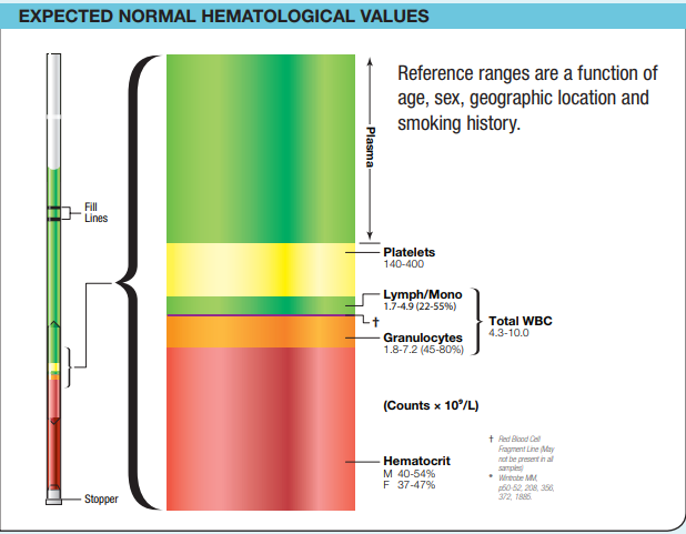
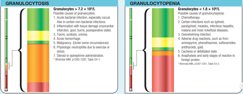
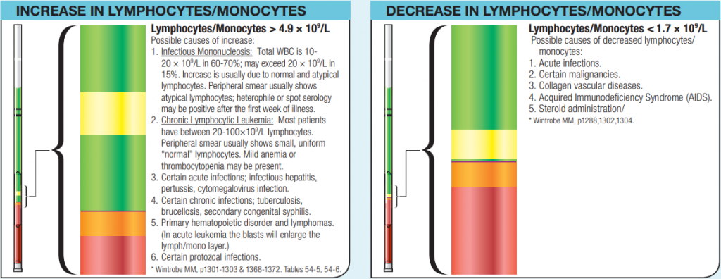
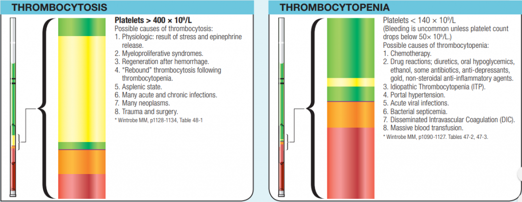
Absolute leukocyte counts, which are obtained by simple multiplication, provide a more accurate and more readily comprehensible picture than the total white blood count with the percentage leukocyte differential count. Although the information is similar in both cases, presenting it in the traditional percentage format may mask abnormalities in the absolute counts.
The importance of the absolute counts is apparent when it is realized that the various leukocyte cell populations are separate entities and under separate control mechanisms; increases in one line may not be paralleled by increases in another. Thus, using the absolute counts as indicators of a particular process offers a more sensitive means than conventional data presentation and also enhances the user's awareness of the pathophysiology.
Note: The QBC system is designed to provide immediate data which can be used as a CBC in many circumstances. The data is not complete, however, and when clinically indicated or when samples show values outside of the approved ranges, the results should be augmented or confirmed by an alternate method, including examination of a stained peripheral blood film.
See package insert and operator's manual. For technical assistance, call 866-265-1486.
- Wintrobe MM, Macroscopic Examination of the Blood, Am J Med Sci 185:58-71, 1933.
- Bessis M. Une Methode Permettant L'isolement des Differents Elements Figures du Sang, Sang 14: 262-264, 1940.
- Davidson E. The Distribution of Cells in the Buffy Layer in Chronic Myeloid Leukemia, Acta Hematologica 23: 22- 28, 1960.
- Zucker RM, Cassen B, The Separation of Normal Human Leucocytes by Density and Classification by Size, Blood 34: 591-600, 1969.
- Wardlaw SC, Levine RA, Quantitative Buffy Coat Analysis, JAMA 249: 617- 620, 1983
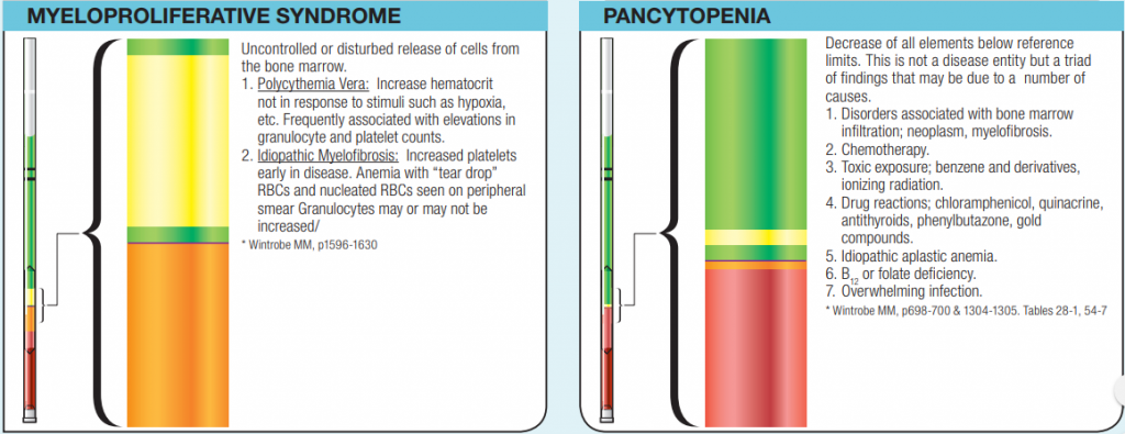
Important observations
Inspection of QBC tube may yield other valuable clinical information.
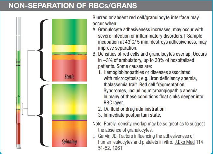
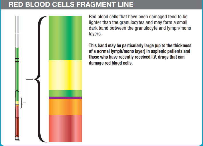
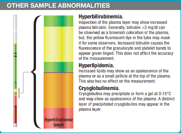
Proper technique ensures accuracy
Inspection of QBC tube prior to reading is an important step in quality assurance.
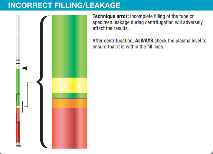
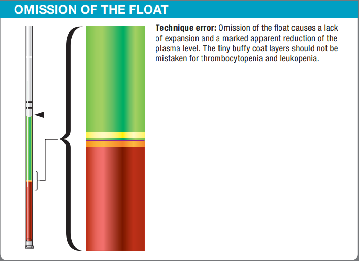
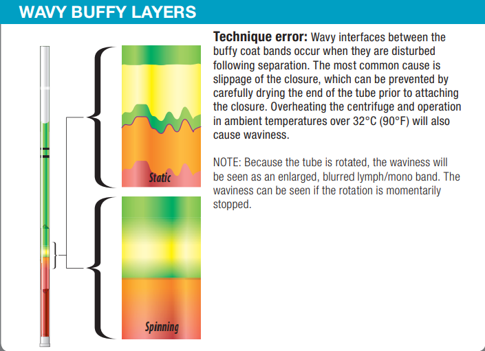
To view the data, visit this link.
Looking for equipment or accessories for your blood testing and other related applications? Feel free to check out our collection of Drucker Diagnostics products at Block Scientific Store today!


